Researchers identify neural plastic alterations that enhance functional recovery after cervical spinal cord injury
Researchers identify neural plastic alterations that enhance functional recovery after cervical spinal cord injury
Researchers at the Indiana University School of Medicine who investigate spinal cord and brain injuries are investigating novel ways to increase functional recovery after a spinal cord injury.
In a recently published article in the Journal of Clinical Investigation, a team of researchers used laboratory models to investigate a unilateral spinal cord injury similar to Brown-Sequard Syndrome, a rare neurological condition characterized by weakness or paralysis on one side of the body and loss of sensation on the opposite side.
Wei Wu, Ph.D., associate research professor of neurological surgery at IU School of Medicine, said that the spinal cord injury model severed the link between the left hemisphere of the brain and the right side of the body, resulting in a severe functional loss in the right forelimb.
"The skilled function of upper limbs is very important for the quality of life in patients with cervical spinal cord injury, but such functional recovery is extremely difficult to achieve in severe injuries," said Wu, the paper's first author and a member of the Spinal Cord and Brain Injury Research Group at Stark Neurological Research Institute.
We discovered that the intact corticospinal system on the opposite side of the brain and spinal cord may be regulated to at least partly take over control of the forelimb that has been injured by a spinal cord injury, resulting in an improvement in forelimb function.
Each hemisphere in the brain controls the opposite side of the body, Wu noted. Researchers identified a spontaneous shift of neuronal networks from the left to the right hemisphere following damage to the left hemisphere. After damage, there exist connections between the right hemisphere of the brain and the right side of the body through certain relayed pathways, but this is insufficient to sustain motor recovery, according to Wu.
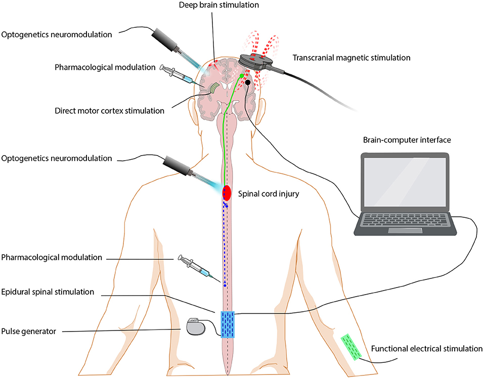
To modify the motor cortex in the right hemisphere of the brain, the scientists used an optogenetic neuromodulation strategy—light stimulation. This transferred new neuronal pathways from the left to the right side of the brain, drastically enhancing and enhancing the function of the forelimbs.
"New circuits are activated in the whisker, mouth, and neck regions of the right hemisphere of the brain to regulate the right forelimb," Wu said.
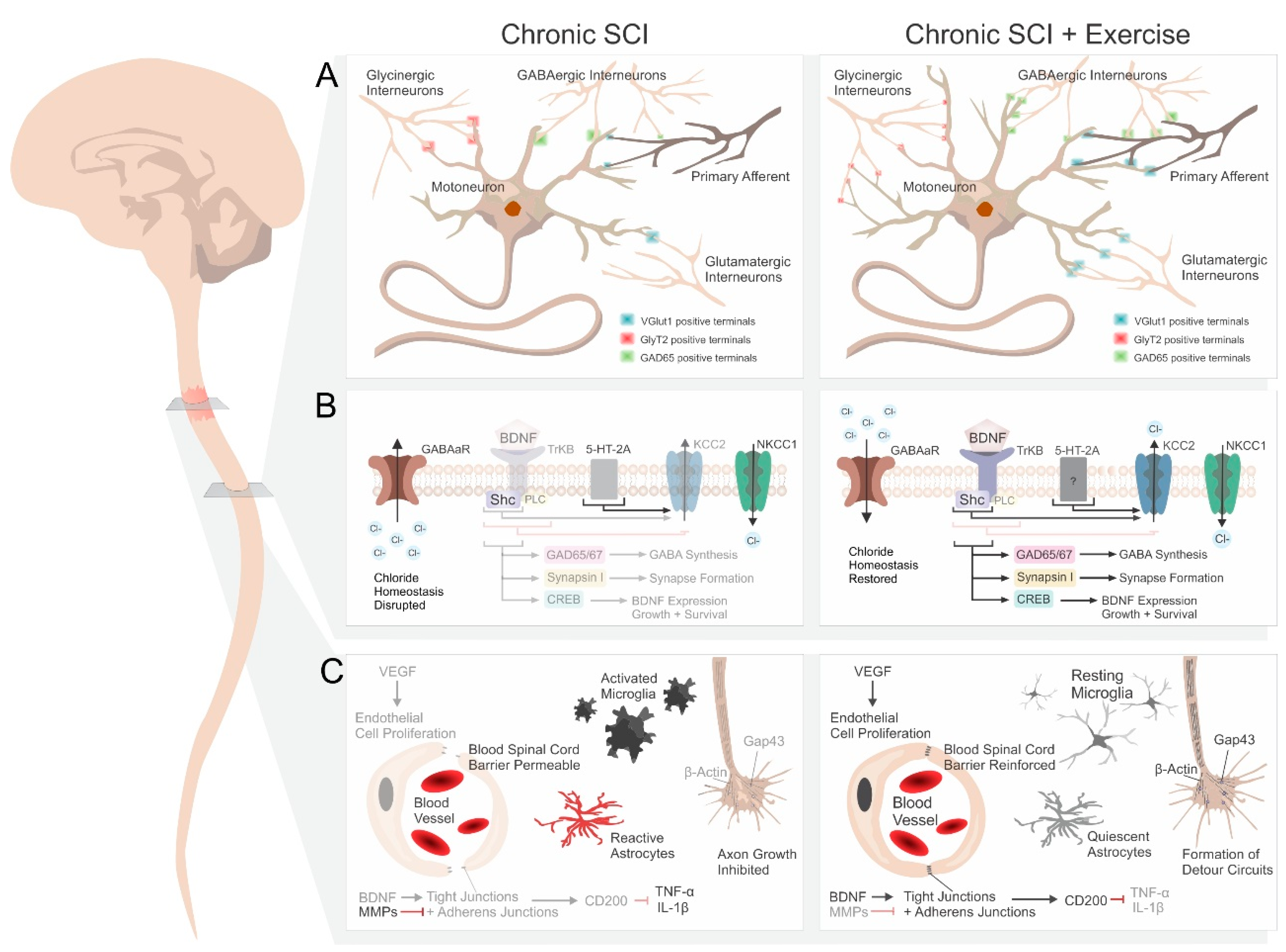
"Intriguingly, when optogenetic neuromodulation was administered to the motor cortex, both the brain and the distal spinal cord exhibited positive neural plastic alterations."
The findings of the research, according to Wu, indicated a considerable improvement in the forelimb; nevertheless, there are still many obstacles to overcome, since total digital recovery has not been achieved.
Wu said that the study team will continue to comprehend and mediate this transhemispheric neuronal remodeling in order to enhance the functional recovery after spinal cord injury.
He anticipates that the findings of this research will be used to the development of a clinical therapy approach for people with spinal cord injury.
Content Source :
https://www.ncbi.nlm.nih.gov/pmc/articles/PMC2562625/


Comments
Post a Comment